
Detail Information of DT Wildtype Structure
| General Information of Drug Transporter (DT) | ||||||
|---|---|---|---|---|---|---|
| DT ID | DTD0534 Transporter Info | |||||
| Gene Name | CACNA1C | |||||
| Transporter Name | Voltage-gated calcium channel alpha Cav1.2 | |||||
| Gene ID | ||||||
| UniProt ID | ||||||
| Modelled Structures of This DT | ||||||
| Click to Save PDB File in TXT Format | ||||||
| Detail Information of Structural Model | ||||||
| Method | Homology modeling | |||||
| Template PDB | 5GJV_A | Identity | 69.088% | |||
| Performance | Minimized Score | -2785.464 kcal/mol | ||||
| Ramachandra Favored |
Medium |
|||||
| QMEANBrane Quality |
Medium |
|||||
| Ramachandran Plot |
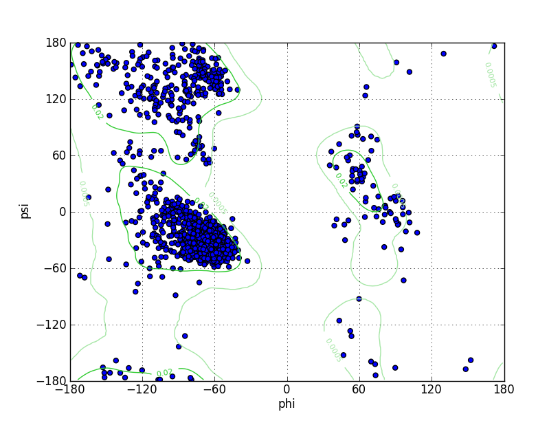
|
|||||
| Click to Save Ramachandran Plot in PNG Format | ||||||
| Ramz Z Score | -2.15 ± 0.18 | |||||
| Residues in Favored Region | 1629 | Ramachandran favored | 95.10% | |||
| Number of Outliers | 18 | Ramachandran outliers | 1.05% | |||
| Local Quality |
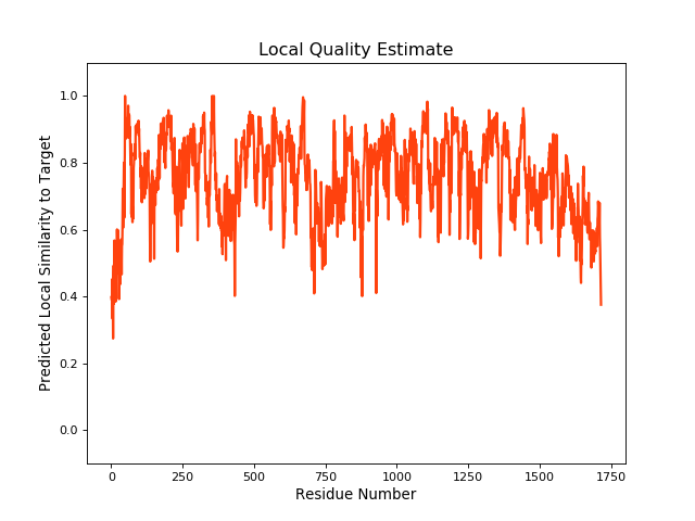
|
|||||
| Click to Save Local Quality Plot in PNG Format | ||||||
| QMEANBrane Score | 0.75 | |||||
| AlphaFold Protein Structure Database | ||||||
| SWISS-MODEL Database | ||||||
| Experimental Structures of This DT | ||||||
| PDB ID | Scanning Method | Resolution | Expression System | Details | Ref | |
| 1T0J | X-ray | 2 Å | Escherichia coli | [1] | ||
| Structure | ||||||
| Click to Save PDB File in TXT Format | ||||||
| Corresponding chain | C | |||||
| Sequence Length | 428-445 | Mutation | No | |||
| 2BE6 | X-ray | 2 Å | Escherichia coli | [2] | ||
| Structure | ||||||
| Click to Save PDB File in TXT Format | ||||||
| Corresponding chain | D/E/F | |||||
| Sequence Length | 1659-1692 | Mutation | No | |||
| 2F3Y | X-ray | 1.45 Å | Escherichia coli BL21(DE3) | [3] | ||
| Structure | ||||||
| Click to Save PDB File in TXT Format | ||||||
| Corresponding chain | B | |||||
| Sequence Length | 1665-1685 | Mutation | No | |||
| 2F3Z | X-ray | 1.6 Å | Escherichia coli BL21(DE3) | [3] | ||
| Structure | ||||||
| Click to Save PDB File in TXT Format | ||||||
| Corresponding chain | B | |||||
| Sequence Length | 1665-1685 | Mutation | Yes | |||
| 2LQC | NMR | N.A. | Escherichia coli | [4] | ||
| Structure | ||||||
| Click to Save PDB File in TXT Format | ||||||
| 3G43 | X-ray | 2.1 Å | Escherichia coli | [5] | ||
| Structure | ||||||
| Click to Save PDB File in TXT Format | ||||||
| Corresponding chain | E/F | |||||
| Sequence Length | 1609-1682 | Mutation | No | |||
| 3OXQ | X-ray | 2.55 Å | Escherichia coli BL21(DE3) | [6] | ||
| Structure | ||||||
| Click to Save PDB File in TXT Format | ||||||
| Corresponding chain | E/F | |||||
| Sequence Length | 1609-1685 | Mutation | No | |||
| 5V2P | X-ray | 2 Å | Escherichia coli | [7] | ||
| Structure | ||||||
| Click to Save PDB File in TXT Format | ||||||
| Corresponding chain | B | |||||
| Sequence Length | 427-445 | Mutation | Yes | |||
| References | ||||||
| 1 | Structure of a complex between a voltage-gated calcium channel beta-subunit and an alpha-subunit domain. Nature. 2004 Jun 10;429(6992):671-5. | |||||
| 2 | Insights into voltage-gated calcium channel regulation from the structure of the CaV1.2 IQ domain-Ca2+/calmodulin complex. Nat Struct Mol Biol. 2005 Dec;12(12):1108-15. | |||||
| 3 | Structure of calmodulin bound to the hydrophobic IQ domain of the cardiac Ca(v)1.2 calcium channel. Structure. 2005 Dec;13(12):1881-6. | |||||
| 4 | Structural basis for the regulation of L-type voltage-gated calcium channels: interactions between the N-terminal cytoplasmic domain and Ca(2+)-calmodulin. Front Mol Neurosci. 2012 Apr 12;5:38. | |||||
| 5 | Crystal structure of dimeric cardiac L-type calcium channel regulatory domains bridged by Ca2+* calmodulins. Proc Natl Acad Sci U S A. 2009 Mar 31;106(13):5135-40. | |||||
| 6 | Multiple C-terminal tail Ca(2+)/CaMs regulate Ca(V)1.2 function but do not mediate channel dimerization. EMBO J. 2010 Dec 1;29(23):3924-38. | |||||
| 7 | Stapled Voltage-Gated Calcium Channel (Ca(V)) -Interaction Domain (AID) Peptides Act As Selective Protein-Protein Interaction Inhibitors of Ca(V) Function. ACS Chem Neurosci. 2017 Jun 21;8(6):1313-1326. | |||||
If you find any error in data or bug in web service, please kindly report it to Dr. Yin and Dr. Li.